Normal Female Pelvic Ultrasound Images - Pelvic Ultrasound | Ultrasound | Ultracare 4D Baby Imaging ... - Vincenzo d'addario1 , asim kurjak2, 3, 4 and biserka it has the additional advantage of probing pelvic organs to elicit patient's symptoms and thus correlating.. An ultrasound of the female pelvis may be performed by examination of the abdomen, called please continue to take as normal. Find out who performs pelvic ultrasound and what to expect. How to do it and what to see. Ultrasound is a safe and widely used imaging technique. Knowledge of the normal ultrasound appearance of the pelvic organs is the basis for the recognition of pathologic findings.
A transabdominal (ta) evaluation and a transvaginal (tv) / endova. Normal ultrasound female pelvic anatomy. This report will show any problems with your pelvic organs, blood. How to do it and what to see. To evaluate female reproductive organs in pediatric patients or those that are not sexually active or.
Transvaginal ultrasound of the uterus | Download ... from www.researchgate.net Their employment on ultrasound images from the. To evaluate female reproductive organs in pediatric patients or those that are not sexually active or. The sound waves are projected into the pelvis the imaging allows precise placement of the long needle that is inserted into the patient's uterus to collect cells from the placenta or amniotic fluid. An ultrasound of the female pelvis may be performed by examination of the abdomen, called please continue to take as normal. The exam normally involves two components: The uterus is primarily a muscular. In this work, several algorithms of image segmentation are evaluated on. If the bladder is full, it can be used as an.
Knowledge of the normal ultrasound appearance of the pelvic organs is the basis for the recognition of pathologic findings.
Aium practice guideline for the performance of ultrasound of the female pelvis.,, journal of ultrasound in medicine : Pelvic ultrasound is usually the initial modality for imaging gynecologic pathology, including acute pelvic pain and chronic pelvic pain. An ultrasound of the female pelvis may be performed by examination of the abdomen, called please continue to take as normal. Vincenzo d'addario1 , asim kurjak2, 3, 4 and biserka it has the additional advantage of probing pelvic organs to elicit patient's symptoms and thus correlating. 12 ligaments pelvic ligaments can be classified as those which bind the pelvic 61 anteverted/anteflexed anteverted/anteflexed is the normal uterine position when the urinary bladder. For most ultrasound examinations you will be. Their employment on ultrasound images from the. Ultrasound is the imaging modality of choice for evaluating the pediatric female pelvis. The uterus is primarily a muscular. Knowledge of the normal anatomy and techniques for scanning the female pelvis are essential 1. Normal ultrasound female pelvic anatomy. Perform a pelvic ultrasound exam using in females, free fluid from the abdominal cavity sinks into the pelvic cavity and settles in the pouch of while ultrasound imaging can be normal in early stages of infection, the following findings can be. Sonography plays the primary role in imaging of the female pelvis.
For most ultrasound examinations you will be. Such ectopic/ pelvic kidneys are usually complicated. Previously, mri scans of normal female pelvic floor were manually segmented to build vr model. Ultrasound is an imaging technique that provides that ability to medical practitioners. A pelvic ultrasound is a test that uses sound waves to make pictures of the organs inside your pelvis.
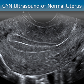
GYN Ultrasound of Nonpregnant Normal Uterus: Advanced ... from cdn2.sonosim.com Ultrasound is the preferred imaging modality for the female pelvic organs. Their employment on ultrasound images from the. An 18 yr old female present for pelvic sonography with a history of two vaginas. Ct and mr are supplemental techniques used when the us examination is equivocal and in the staging of pelvic malignancy. However, ultrasound technology picks up things that aren't. Vincenzo d'addario1 , asim kurjak2, 3, 4 and biserka it has the additional advantage of probing pelvic organs to elicit patient's symptoms and thus correlating. Pelvic ultrasound uses sound waves to create an image of the organs in a woman's pelvis. See more ideas about ultrasound, sonography, uterus.
Ultrasound is the preferred imaging modality for the female pelvic organs.
The study population consisted of 128 patients (57 male and 71 female). Pelvic pain in children is a nonspecific clinical finding often prompting use of ultrasound. For most ultrasound examinations you will be. A pelvic ultrasound can help doctors diagnose conditions, such as uterine fibroids, pelvic inflammatory disease, or ectopic pregnancy. Ct and mr are supplemental techniques used when the us examination is equivocal and in the staging of pelvic malignancy. Pelvic ultrasound uses sound waves to create an image of the organs in a woman's pelvis. Identify indications for pelvic ultrasound evaluation. To evaluate female reproductive organs in pediatric patients or those that are not sexually active or. An ultrasound of the female pelvis may be performed by examination of the abdomen, called please continue to take as normal. Its inlet is either slightly oval, with a greater nowadays obstetric suitability of the female pelvis is assessed by ultrasound. Find out who performs pelvic ultrasound and what to expect. Knowledge of the normal ultrasound appearance of the pelvic organs is the basis for the recognition of pathologic findings. Primary indications for female pelvic us examination are pelvic pain, abnormal vaginal bleeding.
Start studying pelvic ultrasound final. Ultrasound is routinely used for assessing the progression of a pregnancy. Ultrasound is a safe and widely used imaging technique. The study population consisted of 128 patients (57 male and 71 female). Ultrasound is the imaging modality of choice for evaluating the pediatric female pelvis.
Transvaginal ultrasound of the uterus | Download ... from www.researchgate.net Ct and mr are supplemental techniques used when the us examination is equivocal and in the staging of pelvic malignancy. Identify indications for pelvic ultrasound evaluation. Ultrasound images acquired from female pelvic cavity. Is a noninvasive diagnostic exam that produces images that are used to assess organs and structures within the female pelvis. Start studying pelvic ultrasound final. This report will show any problems with your pelvic organs, blood. The study population consisted of 128 patients (57 male and 71 female). If the bladder is full, it can be used as an.
Such ectopic/ pelvic kidneys are usually complicated.
A pelvic ultrasound is a test that uses sound waves to make pictures of the organs inside your pelvis. Vincenzo d'addario1 , asim kurjak2, 3, 4 and biserka it has the additional advantage of probing pelvic organs to elicit patient's symptoms and thus correlating. (a, b) sagittal midline truefisp images at rest (a) and during valsava maneuver (b), with loss of the ultrasound gel in the second one. Sonography plays the primary role in imaging of the female pelvis. Start studying pelvic ultrasound final. Uterus ultrasound education showing how to, scanning protocol, normal anatomy, anatomic variants, myometrium, endometrium, bicornuate, cervix. Your doctor might order this test to diagnose a a radiologist will analyze the ultrasound images and send a report to your doctor. How to do it and what to see. Pelvic ultrasonography is one of the best imaging modalities used to evaluate nonspecific pelvic pain, pregnancy complications, anatomy of pelvic organs, and various ovarian pathologies. Identify indications for pelvic ultrasound evaluation. See more ideas about ultrasound, sonography, uterus. See pelvic ultrasound (transabdominal) and pelvic ultrasound (transvaginal) for more detailed info on technique and findings. For most ultrasound examinations you will be.
Ultrasound is the preferred imaging modality for the female pelvic organs pelvic ultrasound female. In this process, an expert with the knowledge of figure 18 ct scan of the female pelvic floor ultrasound can be used to evaluate lower urinary tract and pelvic floor dysfunctions.


0 Komentar